Advanced Imaging Adaptive Optics & Lens Design
Overview
Simulations Live Labs
Interactive Physics Sandbox
1. Adaptive Optics & PSF Simulator
Zernike modes, turbidity & 3.5x signal boost
2. Ultrafast Pulse & GDD Workbench
Femtosecond chirp, dispersion & 2PEF efficiency
3. Raman 1064 vs 785 nm & SpMap Lab
Autofluorescence & SNIP baseline subtraction
4. Lens Design & Aberration Lab
Bending, spherical & chromatic aberration (2D Optics)
Open Interactive Simulations Lab →
Research & Thesis 10
Publications, Thesis & Theory
Fast Dynamic MEMS Wavefront Shaping
Advanced Imaging (Oct 2026)
2D Optics: Optical Optimization & CAD
Open Source CAD Suite (Oct 2026)
Confocal Raman with Adaptive Optics
ACS Photonics (Dec 2024)
1064 nm Fiber Probe for Bladder Cancer
Applied Sciences (Jul 2024)
Optofluidic SLM in Multi-Photon Imaging
Biomedical Optics Express (2023)
Thesis: AO & Wavefront Shaping
Doctoral Research Pillar
Thesis: Clinical NIR Raman Spectroscopy
Translational Diagnostic Pillar
Theory & Physics Lectures
Raman scattering, multiphoton & chemometrics notes
View all papers & thesis work →
Instruments 6
Optical Systems & Applications
Confocal Raman Microscopy + AO
785 / 532 nm · Confocal pinhole · Optofluidic AO
1064 nm Fiber-Probe Raman
1064 nm NIR · InGaAs array · Clinical ex vivo
Two-Photon Fluorescence (2PEF)
fs Ti:Sa / Fiber · Galvo scanning · DASH & SLM
Coherent Raman (CARS / SRS)
Dual ultrafast · χ⁽³⁾ nonlinear · Video-rate
Photothermal Microscopy
Pump-probe · Lock-in detection · Label-free
Adaptive Optics & Wavefront Shaping
Optofluidic SLM · Zernike modes · Holography
Biomedical & Physical Applications
Tumor diagnostics, neuroscience & deep tissue
Explore all optical instruments →
Software & CAD Apps
Open-Source Packages & App Dev
SpectraMap (GitHub & PyPI)
Hyperspectral clustering, PCA & VCA unmixing
2D Optics (GitHub Workbench)
Lens design, optimization, spectrometers & CAD
Scientific App Development
Instrument control & interactive labs
Explore software & code →
Contact
OverviewSimulationsLive LabsResearch & Thesis10Instruments6Software & CADApps Contact
Juan David Muñoz-Bolaños·Leibniz Institute of Photonic Technology

Advanced Imaging, Adaptive Optics & Lens Design

An interactive scientific portal bridging deep-tissue microscopy, adaptive wavefront shaping, ultrafast optics, and open-source computational toolkits.

⚡ Launch Interactive Simulations 📄 Research Papers & Thesis 💻 SpectraMap & 2D Optics
4 Live Physics Labs 10 Peer-Reviewed & Thesis Works 2 Open-Source Packages 6 Custom Microscopes
Interactive Physics Sandbox

Learn & Experiment with the Physics

Interact with live optical simulations directly in your browser — test tissue scattering, pulse chirps, Raman spectra, and lens aberrations.

Open Full Sandbox (4 Live Labs) →
01 Adaptive Optics

Wavefront Shaping & PSF Recovery

Tissue turbidity scrambles optical phase. An optofluidic SLM or high-speed MEMS on the objective nosepiece cancels aberrations to restore a 3.5× signal boost in mouse brain tissue.

▶ Run AO Simulator (3.5×)
📄 Adv. Imaging (2026) 📄 ACS Phot. (2024)
02 Ultrafast Pulses

Femtosecond Dispersion & GDD

High-NA microscope objectives introduce positive group delay dispersion (GDD), broadening 50 fs pulses and slashing 2PEF efficiency. Prism pre-chirp recovers the transform limit.

▶ Run Pulse Workbench (fs)
👁 2PEF ∝ 1/τ 💻 2D Optics
03 Raman & SpMap

1064 nm Probe & Chemometrics

785 nm laser excitation triggers massive tissue autofluorescence masking tumor peaks. 1064 nm clears background, while SpectraMap automates SNIP baseline subtraction and unmixing.

▶ Run Raman & SpMap Lab
📄 Bladder Resectates 📦 SpMap PyPI
04 Lens Design

Lens Bending & Ray Tracing

Balancing surface curvature (shape factor q = +0.7) eliminates marginal spherical blur, shrinking the spot from 237 µm to diffraction limit. 2D Optics automates DLS optimization.

▶ Run Lens Design Lab
📄 2D Optics Article 💻 GitHub CAD
Peer-Reviewed & Dissertations

Recent Research & Articles

Latest journal publications, doctoral thesis milestones, and scientific instrumentation.

View all (10) articles & thesis →
Fast Dynamic Wavefront Shaping for Multiphoton Microscopy using Piston-like MEMs Adaptive Optics
Advanced Imaging (2026) Oct 27, 2026

Fast Dynamic Wavefront Shaping for Multiphoton Microscopy using Piston-like MEMs

Sensorless phase retrieval leveraging a fast high-resolution MEMS phase-only modulator targeting real-time aberration correction in nonlinear multiphoton microscopy across organoids and brain tissue.
Read paper → AO & Wavefront Simulator →
2D Optics: Zemax-Standard Optical Optimization, Laser Resonators & AI Engineering Optical CAD & AI
Open Source CAD Suite Oct 27, 2026

2D Optics: Zemax-Standard Optical Optimization, Laser Resonators & AI Engineering

Desktop optical engineering suite featuring a full Lens Data Editor (LDE), sequential/non-sequential ray tracing, Zemax-standard LM-DLS optimizer, laser cavity stability, 54 Herbert Gross presets, and STEP/DXF CAD export.

Read paper → Lens Design & Aberration Lab →
Confocal Raman Microscopy with Adaptive Optics Adaptive Optics
ACS Photonics (2024) Dec 20, 2024

Confocal Raman Microscopy with Adaptive Optics

First application of adaptive optics in confocal Raman microscopy: a transmissive optofluidic wavefront shaper on the objective nosepiece corrects sample-induced aberrations, boosting Raman signal up to 3.5x in mouse brain tissue.

Read paper → AO & Wavefront Simulator →
Open-Source Toolkits

Scientific Software & CAD Workbenches

Production software for hyperspectral chemometrics and Zemax-standard optical engineering.

Explore Software & Manuals →
SpectraMap (SpMap)
PyPI · MIT License Python 3 · Streamlit

SpectraMap (SpMap)

High-throughput hyperspectral analysis, batch parallel chemometrics & Streamlit GUI in Python

SpectraMap is a validated Python framework covering the entire hyperspectral workflow: parallel batch loading, automated wavenumber resampling, SNIP/airPLS baselines, internal standard peak normalization, parallel PCA & HCA, HDBSCAN clustering, and VCA unmixing. Includes a 5-step guided Streamlit web application with colorblind-friendly overlays, ROI map manipulation, and publication figure export.

Features & gallery → GitHub ↗PyPI ↗Manual ↗Blog postRaman & SpMap Baseline Lab →
2D Optics
GitHub · Desktop App Python · Qt · pyqtgraph

2D Optics

Zemax-standard optical optimization, laser cavities, 54 Herbert Gross presets & AI workbench

2D Optics is a desktop optical engineering suite featuring a full Lens Data Editor (LDE), sequential and non-sequential 2D/3D ray tracing, and a Zemax-standard DLS Levenberg-Marquardt optimizer. It models complete instruments (Raman spectrometers, multiphoton objectives, laser cavities, telecentric relays, light-pipe homogenizers) and exports manufacturing-ready CAD.

Features & gallery → GitHub ↗Blog postLens Design & Aberration Lab →
Interactive Physics Sandbox

Live Optical & Spectroscopic Simulations

Explore the physics behind our microscopes in real time: adjust Zernike wavefront aberrations, femtosecond group delay dispersion (GDD), and Raman excitation wavelengths directly in your browser.

1. Adaptive Optics 2. Ultrafast Pulses 3. Raman & SpMap 4. Lens Design
Simulator 01 · Wavefront Shaping & Point Spread Function

Adaptive Optics & Nosepiece Wavefront Correction

Modality:
💡 Try This Experiment Observe: Scrambled tissue phase scatters the focus, dropping Strehl ratio to <0.20. Action: Click Mouse Brain (3.5×) to see the optofluidic SLM restore a sharp diffraction-limited spot!
Strehl S → Raman I ∝ S · 2PEF I ∝ S²
Tissue Scattering / Turbidity (σϕ) 1.4 rad
Spherical Aberration (Z40) 0.8 rad
Astigmatism & Coma (Z22) -0.6 rad
Optofluidic SLM / AO Correction 85%
Residual Pupil Phase Δϕ(x,y)
RMS: 0.28 rad
2D Focal Spot PSF I(x,y)
Strehl: 0.78
Lateral Focus Profile I(x)
Signal Boost: 3.4×
Simulator 02 · 2D Optics Ultrafast Workbench

Femtosecond Pulse Dispersion (GDD) & Nonlinear Efficiency

💡 Try This Experiment Observe: High-NA glass dispersion (+1800 fs²) stretches a 50 fs pulse to >61 fs. Action: Click Prism Pre-Chirp (0 net fs²) to introduce negative dispersion and compress the pulse at the sample!
τout = τ0√(1 + (4 ln2 GDD/τ0²)²)
Transform-Limited Pulse (τ0) 50 fs
Optical Glass / Objective GDD +1800 fs²
Compressor Pre-Chirp (GDDcomp) -1200 fs²
Sample Pulse τ
61.6 fs
2PEF / CARS Signal
81.2%
Temporal Envelope I(t) & Chirped Carrier E(t) Net GDD: +600 fs² · Δλ @ 800 nm: 18.8 nm
Simulator 03 · Clinical Raman & SpMap Processing

785 nm vs. 1064 nm Excitation & SNIP Baseline Removal

💡 Try This Experiment Observe: 785 nm creates a massive autofluorescence background in bladder tissue. Action: Click 1064 nm Fiber Probe, then toggle SpMap SNIP Baseline to see the unmixed collagen & protein peaks!
SpMap SNIP Baseline Subtraction
Laser Excitation Wavelength
Integration Time / Signal-to-Noise High (80%)
Key bands: 855, 1003 (Phe), 1260 (Amide III), 1445 (CH₂), 1655 (Amide I) cm⁻¹
Raman Shift Spectrum (400 – 1800 cm⁻¹) ── Raw + Autofluorescence   ━━ SpMap Unmixed Raman
Simulator 04 · 2D Optics Exact Ray Trace

Lens Design: Lens Bending, Spherical & Chromatic Aberration

💡 Try This Experiment Observe: Plano-convex flat side first (q = -1) has massive spherical aberration (237 µm spot). Action: Click Best-form singlet (q = +0.70) to shrink the spot to ~36 µm, then click Find best focus!
Shape factor q = (R₂+R₁)/(R₂−R₁)
Lens bending (shape factor q) +0.70
Aperture (f-number, f = 100 mm) f/4.0
Image-plane defocus 0.00 mm
Glass
RMS spot
—
Airy radius
—
Meridional ray trace (exact Snell refraction at spherical surfaces) R₁ · R₂
Transverse ray fan εy(ρ) at image plane
RMS spot radius vs. focus position

Longitudinal spherical aberration: —

Optical Instrumentation

Imaging Microscopes

Custom-built optical microscopes and spectroscopic imaging systems combining spontaneous/coherent Raman, two-photon fluorescence, and adaptive wavefront control.

Confocal Raman Microscopy + AO λ

Confocal Raman Microscopy + AO

Label-free chemical imaging with diffraction-limited 3D sectioning. Integrates a sensorless optofluidic phase plate directly on the objective nosepiece.

785 / 532 nmConfocal pinholeOptofluidic AO
  • 📖 Raman Imaging
  • 📄 Confocal Raman Microscopy with Adaptive Optics
  • 📄 Raman Intensity Calibration from 500 to 1100 nm Laser Excitation
  • 📄 Raman Wavenumber Calibration by Paracetamol (785 nm)
AO & Wavefront Simulator →
1064 nm Fiber-Probe Raman ∿

1064 nm Fiber-Probe Raman

Dispersive InGaAs NIR Raman spectrometer coupled to a flexible fiber probe. 1064 nm excitation suppresses biological autofluorescence in clinical tissues.

1064 nm NIRInGaAs arrayClinical ex vivo
  • 📖 Raman Imaging
  • 📄 1064 nm Fiber Probe Raman Imaging for Human Bladder Resectates
Raman & SpMap Baseline Lab →
Two-Photon Fluorescence (2PEF) 2γ

Two-Photon Fluorescence (2PEF)

Femtosecond laser-scanning microscopy with intrinsic 3D sectioning and deep tissue penetration, enhanced with holographic & optofluidic wavefront control.

fs Ti:Sa / FiberGalvo scanningDASH & SLM
  • 📖 Two-Photon Excitation Fluorescence (2PEF)
  • 📄 TPEF: Improving Speed and Deep Tissue Imaging by Holography (DASH & PMC)
  • 📄 Optofluidic Spatial Light Modulator in Multi-Photon Imaging
Ultrafast Pulse & GDD Simulator →
Coherent Raman (CARS / SRS) ω

Coherent Raman (CARS / SRS)

Synchronized pump and Stokes femtosecond/picosecond pulse trains coherently drive molecular vibrations for video-rate chemical microscopy.

Dual ultrafastχ⁽³⁾ nonlinearVideo-rate
  • 📖 Coherent Raman Scattering (CARS / SRS)
Ultrafast Pulse & GDD Simulator →
ΔT
ΔT

Photothermal Microscopy

Modulated pump beam heats absorbing targets locally; a co-propagating probe beam detects the resulting thermal lens refractive-index modulation.

Pump-probeLock-in detectionLabel-free
  • 📖 Photothermal Microscopy
Adaptive Optics & Wavefront Shaping ≈

Adaptive Optics & Wavefront Shaping

Refractive optofluidic SLMs and holographic algorithms (DASH, PMC) that measure and pre-compensate phase aberrations and multiple scattering.

Optofluidic SLMZernike modesHolography
  • 📖 Two-Photon Excitation Fluorescence (2PEF)
  • 📄 Confocal Raman Microscopy with Adaptive Optics
  • 📄 Optofluidic Spatial Light Modulator in Multi-Photon Imaging
  • 📄 TPEF: Improving Speed and Deep Tissue Imaging by Holography (DASH & PMC)
AO & Wavefront Simulator →
Translational Research

Biomedical & Physical Applications

Clinical tumor diagnostics, neuroscience aberration correction, deep-tissue imaging, urology, spectroscopic metrology, and hyperspectral data science.

Oncology & Bladder Tumor Diagnostics

Oncology & Bladder Tumor Diagnostics

Label-free 1064 nm Raman imaging discriminates tumor, necrosis and healthy urothelium in human bladder resectates.

  • 📖 Machine Learning for Hyperspectral Images
  • 📄 1064 nm Fiber Probe Raman Imaging for Human Bladder Resectates
Neuroscience & Deep Brain Tissue

Neuroscience & Deep Brain Tissue

Sensorless adaptive optics corrects myelinated tissue aberrations in mouse brain, recovering spatial resolution and 3.5x chemical signal.

  • 📖 Raman Imaging
  • 📄 Confocal Raman Microscopy with Adaptive Optics
Turbid & Scattering Media Imaging

Turbid & Scattering Media Imaging

Holographic wavefront shaping (DASH/PMC) and transmissive SLMs form sharp foci through strongly scattering biological layers.

  • 📖 Two-Photon Excitation Fluorescence (2PEF)
  • 📄 TPEF: Improving Speed and Deep Tissue Imaging by Holography (DASH & PMC)
  • 📄 Optofluidic Spatial Light Modulator in Multi-Photon Imaging
Urology & Kidney Stone Analysis

Urology & Kidney Stone Analysis

Rapid mineralogical identification of highly fluorescent kidney stones where conventional 785 nm Raman spectrometers fail.

  • 📄 1064 nm Fiber Probe Raman Imaging for Human Bladder Resectates
Spectroscopic Metrology & Calibration

Spectroscopic Metrology & Calibration

Standardized wavenumber (paracetamol) and broad-spectrum intensity calibration protocols for cross-instrument reproducibility.

  • 📄 Raman Wavenumber Calibration by Paracetamol (785 nm)
  • 📄 Raman Intensity Calibration from 500 to 1100 nm Laser Excitation
Hyperspectral Chemometrics & AI

Hyperspectral Chemometrics & AI

Automated spectral preprocessing, cosmic-ray removal, clustering, and NMF unmixing implemented in the SpMap Python library.

  • 📖 Machine Learning for Hyperspectral Images
  • 📄 SpMap: Hyperspectral Package for Spectroscopists
  • 📄 PDF LaTeX Equations to LaTeX Code by Deep Learning (pix2tex)
Foundations

Theory & Lecture Notes

Concise physical derivations and instrumentation notes on Raman scattering, coherent nonlinear optics, multiphoton excitation, photothermal contrast, and chemometrics.

Vibrational Physics

Raman Imaging

Inelastic light scattering, molecular polarizability, vibrational modes, and how hyperspectral Raman cubes are acquired and unmixed into chemical maps.

Read lecture → Raman & SpMap Baseline Lab →
Nonlinear Optics

Coherent Raman Scattering (CARS / SRS)

Third-order nonlinear susceptibility χ⁽³⁾, coherent driving of molecular vibrations with pump and Stokes ultrafast pulses, and video-rate chemical imaging.

Read lecture → Ultrafast Pulse & GDD Simulator →
Multiphoton Physics

Two-Photon Excitation Fluorescence (2PEF)

Quadratic intensity dependence, intrinsic 3D optical sectioning, NIR scattering reduction, and femtosecond pulse duration requirements.

Read lecture → Ultrafast Pulse & GDD Simulator →
Absorption Contrast

Photothermal Microscopy

Pump-probe detection of local refractive-index gradients (∂n/∂T) induced by optical absorption in non-fluorescent nanostructures and tissues.

Read lecture →
Data Science

Machine Learning for Hyperspectral Images

Dimensionality reduction (PCA), k-means/hierarchical clustering, NMF/vertex spectral unmixing, and supervised tissue classification.

Read lecture → Raman & SpMap Baseline Lab →

Calibration Protocols & Computational Methods

TPEF: Improving Speed and Deep Tissue Imaging by Holography (DASH & PMC) Wavefront Shaping
Jan 16, 2022 Jan 16, 2022

TPEF: Improving Speed and Deep Tissue Imaging by Holography (DASH & PMC)

Dynamic Adaptive Scattering compensation Holography (DASH) and phase-modulation methods that recover diffraction-limited two-photon focus deep inside turbid tissue.

Read paper → AO & Wavefront Simulator →
Raman Intensity Calibration from 500 to 1100 nm Laser Excitation Calibration
Mar 5, 2022 Mar 5, 2022

Raman Intensity Calibration from 500 to 1100 nm Laser Excitation

Standardized protocol to calibrate the spectral intensity (y-axis) response of Raman spectrometers across visible and NIR excitation wavelengths.

Read paper → Raman & SpMap Baseline Lab →
Raman Wavenumber Calibration by Paracetamol (785 nm) Calibration
Jan 12, 2022 Jan 12, 2022

Raman Wavenumber Calibration by Paracetamol (785 nm)

Wavenumber (x-axis) calibration at 785 nm using paracetamol reference vibrational peaks and polynomial pixel-to-wavenumber fitting.

Read paper → Raman & SpMap Baseline Lab →
PDF LaTeX Equations to LaTeX Code by Deep Learning (pix2tex) Machine Learning
Jan 17, 2022 Jan 17, 2022

PDF LaTeX Equations to LaTeX Code by Deep Learning (pix2tex)

Convert printed scientific equations from PDF papers into editable LaTeX source code using vision-transformer deep learning.

Read paper →
Academic Portfolio

Research Papers, Thesis Work & Technical Notes

Peer-reviewed journal publications, doctoral thesis research pillars, and calibration tutorials. Any new post published to Blogger appears here automatically.

Thesis & Dissertation Research Pillars

Doctoral & Advanced Research Program 2021 – 2025

Adaptive Optics & Wavefront Shaping for Deep-Tissue Chemical and Nonlinear Microscopy

Investigating transmissive optofluidic spatial light modulators, sensorless modal wavefront optimization, and holographic scattering compensation (DASH/PMC) to overcome sample-induced aberrations in spontaneous confocal Raman and multiphoton microscopy.

  • ·Nosepiece integration of a 2D transmissive optofluidic phase modulator into a confocal Raman microscope (3.5x signal recovery in mouse brain).
  • ·Low-loss refractive wavefront shaping for two-photon excitation fluorescence (2PEF) without complex folded relay optics.
  • ·End-to-end calibration, spectral unmixing (SpMap), and ray-tracing/ultrafast optical design (2D Optics).
Associated Publications & Interactive Model
📄 Fast Dynamic Wavefront Shaping for Mul…📄 Confocal Raman Microscopy with Adaptiv…📄 Optofluidic Spatial Light Modulator in…📄 TPEF: Improving Speed and Deep Tissue … AO & Wavefront Simulator →
Biomedical Spectroscopy & Instrumentation 2022 – 2024

Translational NIR Fiber-Probe Raman Spectroscopy for Clinical Diagnostics

Design, calibration, and clinical validation of a compact 1064 nm dispersive fiber-probe Raman imaging system for label-free tumor margin assessment in human bladder resectates and urological stone analysis.

  • ·1064 nm NIR excitation eliminates tissue autofluorescence that overwhelms 785 nm spectra in bladder and kidney specimens.
  • ·Automated hyperspectral preprocessing, SNIP baseline subtraction, PCA, and clustering via the SpMap Python library.
  • ·NIST-traceable wavenumber and white-light intensity calibration across 500–1100 nm.
Associated Publications & Interactive Model
📄 1064 nm Fiber Probe Raman Imaging for …📄 SpMap: Hyperspectral Package for Spect…📄 Raman Intensity Calibration from 500 t…📄 Raman Wavenumber Calibration by Parace… Raman & SpMap Baseline Lab →

Published Articles & Technical Posts

Fast Dynamic Wavefront Shaping for Multiphoton Microscopy using Piston-like MEMs Adaptive Optics
Advanced Imaging (2026) Oct 27, 2026

Fast Dynamic Wavefront Shaping for Multiphoton Microscopy using Piston-like MEMs

Sensorless phase retrieval leveraging a fast high-resolution MEMS phase-only modulator targeting real-time aberration correction in nonlinear multiphoton microscopy across organoids and brain tissue.
Read paper → AO & Wavefront Simulator →
2D Optics: Zemax-Standard Optical Optimization, Laser Resonators & AI Engineering Optical CAD & AI
Open Source CAD Suite Oct 27, 2026

2D Optics: Zemax-Standard Optical Optimization, Laser Resonators & AI Engineering

Desktop optical engineering suite featuring a full Lens Data Editor (LDE), sequential/non-sequential ray tracing, Zemax-standard LM-DLS optimizer, laser cavity stability, 54 Herbert Gross presets, and STEP/DXF CAD export.

Read paper → Lens Design & Aberration Lab →
Confocal Raman Microscopy with Adaptive Optics Adaptive Optics
ACS Photonics (2024) Dec 20, 2024

Confocal Raman Microscopy with Adaptive Optics

First application of adaptive optics in confocal Raman microscopy: a transmissive optofluidic wavefront shaper on the objective nosepiece corrects sample-induced aberrations, boosting Raman signal up to 3.5x in mouse brain tissue.

Read paper → AO & Wavefront Simulator →
1064 nm Fiber Probe Raman Imaging for Human Bladder Resectates Clinical Raman
Applied Sciences (2024) Jul 7, 2024

1064 nm Fiber Probe Raman Imaging for Human Bladder Resectates

Compact dispersive 1064 nm fiber-probe Raman imaging spectrometer that suppresses biological autofluorescence, discriminates tumor from non-tumor human bladder tissue and characterizes kidney stones.

Read paper → Raman & SpMap Baseline Lab →
Optofluidic Spatial Light Modulator in Multi-Photon Imaging Multiphoton AO
Biomedical Optics Express (2023) Feb 10, 2023

Optofluidic Spatial Light Modulator in Multi-Photon Imaging

Transmissive, low-loss optofluidic phase modulator for sensorless aberration correction in two-photon excitation fluorescence microscopy.

Read paper → AO & Wavefront Simulator →
SpMap: Hyperspectral Package for Spectroscopists Software
PyPI Open Source Aug 15, 2022

SpMap: Hyperspectral Package for Spectroscopists

Open-source Python 3 package for hyperspectral images: preprocessing, baseline correction, PCA, clustering and spectral unmixing for Raman, IR and fluorescence data.

Read paper → Raman & SpMap Baseline Lab →
Raman Intensity Calibration from 500 to 1100 nm Laser Excitation Calibration
Mar 5, 2022 Mar 5, 2022

Raman Intensity Calibration from 500 to 1100 nm Laser Excitation

Standardized protocol to calibrate the spectral intensity (y-axis) response of Raman spectrometers across visible and NIR excitation wavelengths.

Read paper → Raman & SpMap Baseline Lab →
PDF LaTeX Equations to LaTeX Code by Deep Learning (pix2tex) Machine Learning
Jan 17, 2022 Jan 17, 2022

PDF LaTeX Equations to LaTeX Code by Deep Learning (pix2tex)

Convert printed scientific equations from PDF papers into editable LaTeX source code using vision-transformer deep learning.

Read paper →
TPEF: Improving Speed and Deep Tissue Imaging by Holography (DASH & PMC) Wavefront Shaping
Jan 16, 2022 Jan 16, 2022

TPEF: Improving Speed and Deep Tissue Imaging by Holography (DASH & PMC)

Dynamic Adaptive Scattering compensation Holography (DASH) and phase-modulation methods that recover diffraction-limited two-photon focus deep inside turbid tissue.

Read paper → AO & Wavefront Simulator →
Raman Wavenumber Calibration by Paracetamol (785 nm) Calibration
Jan 12, 2022 Jan 12, 2022

Raman Wavenumber Calibration by Paracetamol (785 nm)

Wavenumber (x-axis) calibration at 785 nm using paracetamol reference vibrational peaks and polynomial pixel-to-wavenumber fitting.

Read paper → Raman & SpMap Baseline Lab →
Open-Source Ecosystem

Scientific Software & Application Development

SpectraMap for hyperspectral chemometrics and 2D Optics for lens design, spectrometer engineering and ultrafast optics — both developed in the lab and published on GitHub.

SpectraMap 2D Optics App Development
SpectraMap (SpMap)
PyPI · MIT License Python 3 · Streamlit

SpectraMap (SpMap)

High-throughput hyperspectral analysis, batch parallel chemometrics & Streamlit GUI in Python

SpectraMap is a validated Python framework covering the entire hyperspectral workflow: parallel batch loading, automated wavenumber resampling, SNIP/airPLS baselines, internal standard peak normalization, parallel PCA & HCA, HDBSCAN clustering, and VCA unmixing. Includes a 5-step guided Streamlit web application with colorblind-friendly overlays, ROI map manipulation, and publication figure export.

Parallel
batch engine
5-Step
Streamlit GUI
100+
unit tests
MIT / PyPI
open source
pip install spectramap
GitHub ↗PyPI ↗Manual ↗Blog postRaman & SpMap Baseline Lab →
High-Performance Breakthroughs & Latest Updates
⚡ Parallel Batch Engine
Parallel multi-file loading via joblib, automatic linear wavenumber resampling onto a common grid, and accelerated IncrementalPCA.
🎯 Internal Standard Normalization
Locks intensities against known vibrational reference bands (e.g. Paracetamol, Silicon, Amide I) to cancel laser fluctuation and focal drift.
🎨 5-Step Guided Stepper GUI
Interactive Streamlit UI with Okabe-Ito (colorblind-safe) and CMY/CMYW channel overlays, map rotate/flip/crop, and automated figure export.
Capabilities
High-Throughput Batch Processing
Parallel batch dataset loading via joblib, automatic linear wavenumber resampling onto a common grid, and group difference spectra with sample classification breakdown.
Internal Standard Peak Normalization
Normalize intensities against designated vibrational standard peaks (e.g. paracetamol, silicon, collagen) to cancel cross-measurement baseline and instrument drift.
Validated Data Type Architecture
Strict type enforcement across hyper_image, multi_spectra, and single_spectrum objects to prevent invalid spatial/spectral operations.
Advanced Baseline & Preprocessing
Spike removal (modified z-score), fast SNIP, rubberband, and airPLS baseline correction, Savitzky-Golay and Gaussian smoothing, vector and area normalization.
Clustering & Spatial Segmentation
K-means, parallelized Hierarchical Cluster Analysis (run_hca_parallel), and density-based HDBSCAN segmentation with cluster-averaged spectral stacks and scatter plots.
Chemometrics & Unmixing
Parallel IncrementalPCA with score scatter profiling, PLS-LDA classification, and Vertex Component Analysis (VCA) for pure endmember extraction with global sync.
5-Step Guided Streamlit Stepper
Interactive GUI (app.py) with Okabe-Ito (colorblind-safe) and CMY/CMYW channel overlays, map rotate/flip/crop, and one-click export of figures, tables, and JSON metadata.
Smart AI Importer & Flexible I/O
Ollama/Nemotron LLM-assisted parser for custom headers, plus native readers for CSV, TXT, Thermo/Galactic SPC (single/multi/holographic), and MATLAB .mat files.
Gallery (from the GitHub repository)
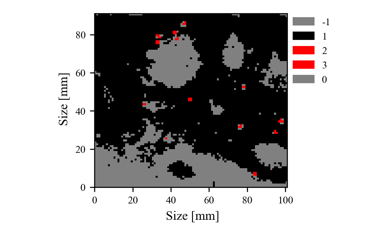
HDBSCAN segmentation of microplastics in tissue
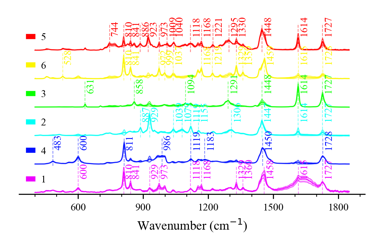
Cluster-averaged spectral stack
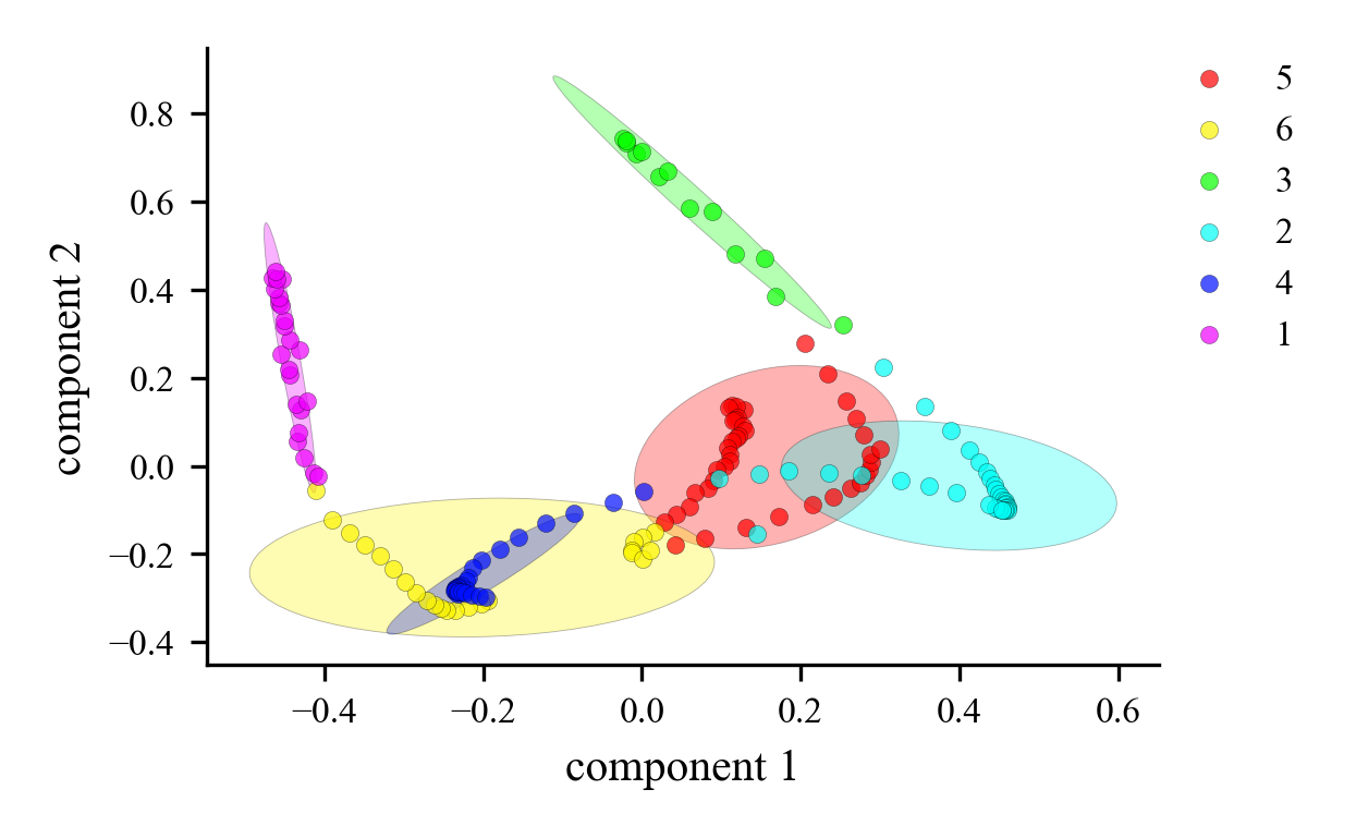
PCA scores separating biomolecular components
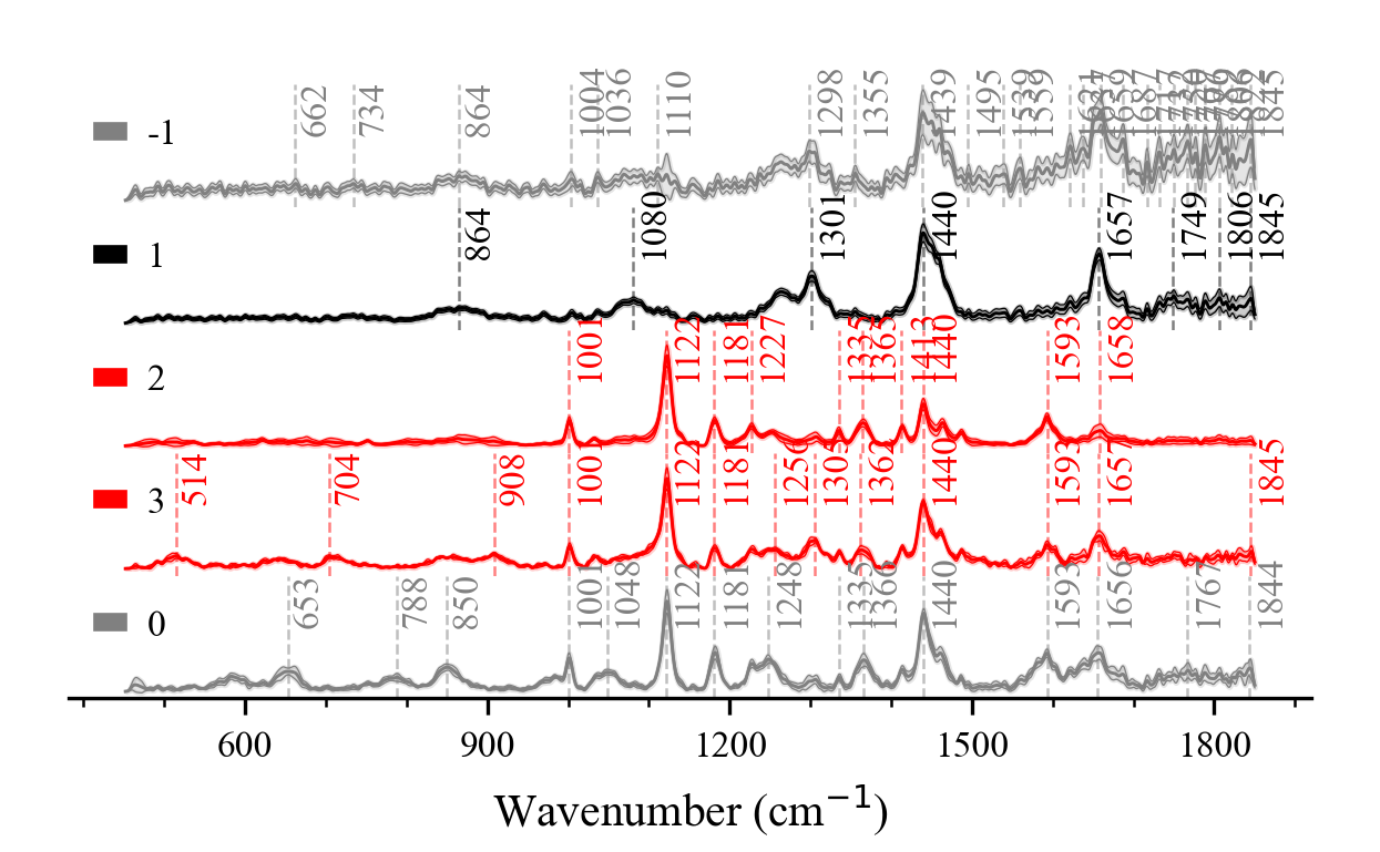
Mean ± SD Raman signature of tissue
Quick start
from spectramap import spmap as sp

micro = sp.hyper_object('Microplastics', data_type='hyper_image')
micro.read_csv_xz('examples/microplastics_tissue/microplastics_tissue')
micro.keep(400, 1850)   # fingerprint region
micro.snip(30)          # SNIP baseline
micro.gaussian(2)
micro.vector()
micro.hdbscan(5, 5)     # density clustering
colors = micro.show_map(['gray', 'k', 'r'], None, 1)
micro.show_stack(0.1, 0.5, colors)
2D Optics
GitHub · Desktop App Python · Qt · pyqtgraph

2D Optics

Zemax-standard optical optimization, laser cavities, 54 Herbert Gross presets & AI workbench

2D Optics is a desktop optical engineering suite featuring a full Lens Data Editor (LDE), sequential and non-sequential 2D/3D ray tracing, and a Zemax-standard DLS Levenberg-Marquardt optimizer. It models complete instruments (Raman spectrometers, multiphoton objectives, laser cavities, telecentric relays, light-pipe homogenizers) and exports manufacturing-ready CAD.

54
Gross presets
Zemax DLS
LM optimizer
2,650+
verified tests
ISO 10110
manufacturing audit
git clone https://github.com/MicroscopeMaestro/2D_Optics.git
GitHub ↗Blog postLens Design & Aberration Lab →
Zemax Parity & Optical Engineering Breakthroughs
📐 Zemax LM-DLS & Hammer Optimizer
Multi-field area weighting, hexapolar & Gauss-Legendre pupil sampling, Rayleigh-Hamilton OPD targets, and boundary operands (MNCT, MXCT, MNET, MNEA).
🔬 Laser Resonators & Homogenizers
ABCD matrix stability for Ti:Sapphire Z-fold & Nd:YAG thermal cavities, tandem fly's-eye MLA homogenizers, and double-telecentric mask projection.
🤖 54 Gross Presets & AI Explainer
Digitized Volumes 1–6 of the Handbook of Optical Systems, Nemotron AI optical assistant, and ISO 10110 manufacturing audit with STEP/DXF export.
Capabilities
Zemax-Standard LM-DLS Engine
Native multi-surface Levenberg-Marquardt Damped Least Squares optimizer with adaptive damping (lambda_{k+1} = lambda_k * beta), SVD/Cholesky conditioning, and step backtracking; Cauchy/Gaussian Hammer simulated annealing.
Multi-Field & Pupil Quadrature
Standard Hy in [0.0, 0.707, 1.0] with area weighting [1, 2, 1], full-pupil hexapolar up to rho=1.0, Gauss-Legendre quadrature grids, and paraxial Rayleigh-Hamilton RMS wavefront OPD scaling.
Zemax Boundary Operands
Numerical barrier penalties for MNCT, MXCT (1.5 to 18 mm), MNET, MXET (ET >= 1.0 mm at clear semi-diameter via exact sag), MNEA, EFFL (paraxial power), and TOTR (Surface 1 to image).
54 Digitized Herbert Gross Presets
Volumes 1 to 6 of the Handbook of Optical Systems: Cooke triplets, Double Gauss 50mm f/2, Petzval portrait, Zeiss Tessar, Klevtsov, Schmidt-Cassegrain, Offner and Dyson 1x relays, and zoom lenses.
Laser Cavity Resonators & Stability
ABCD matrix round-trip stability for Ti:Sapphire astigmatic-compensated Z-fold cavities (800 nm), Bowtie ring resonators, and Nd:YAG thermal lens cavities (1064 nm).
Homogenizers & Mask Projection
Tandem fly's-eye microlens arrays (MLA) and TIR kaleidoscope light pipes for laser beam homogenization; double-telecentric reticle/mask projection systems.
Ultrafast & Nonlinear Imaging
Femtosecond pulse dispersion (GDD/TOD per ray path), two-photon fluorescence (2PEF), and calibrated CARS/SRS/SFG/DFG nonlinear imaging workbench with PMT models.
AI Optical Assistant & ISO 10110
Nemotron LLM-assisted generative design sequences, automated aberration diagnosis, and ISO 10110 manufacturing report with zero-violation verification and STEP/DXF/SVG export.
Gallery (from the GitHub repository)
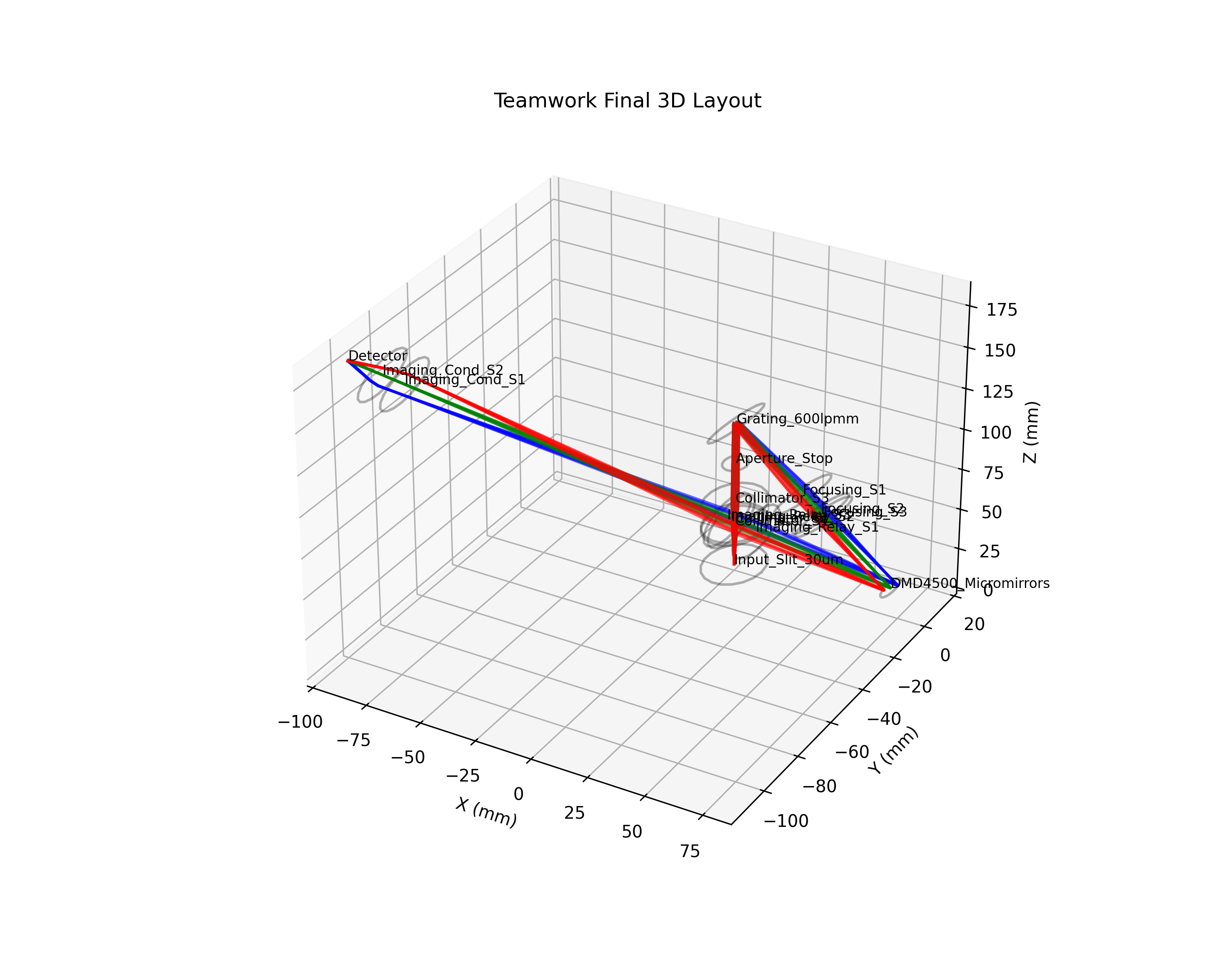
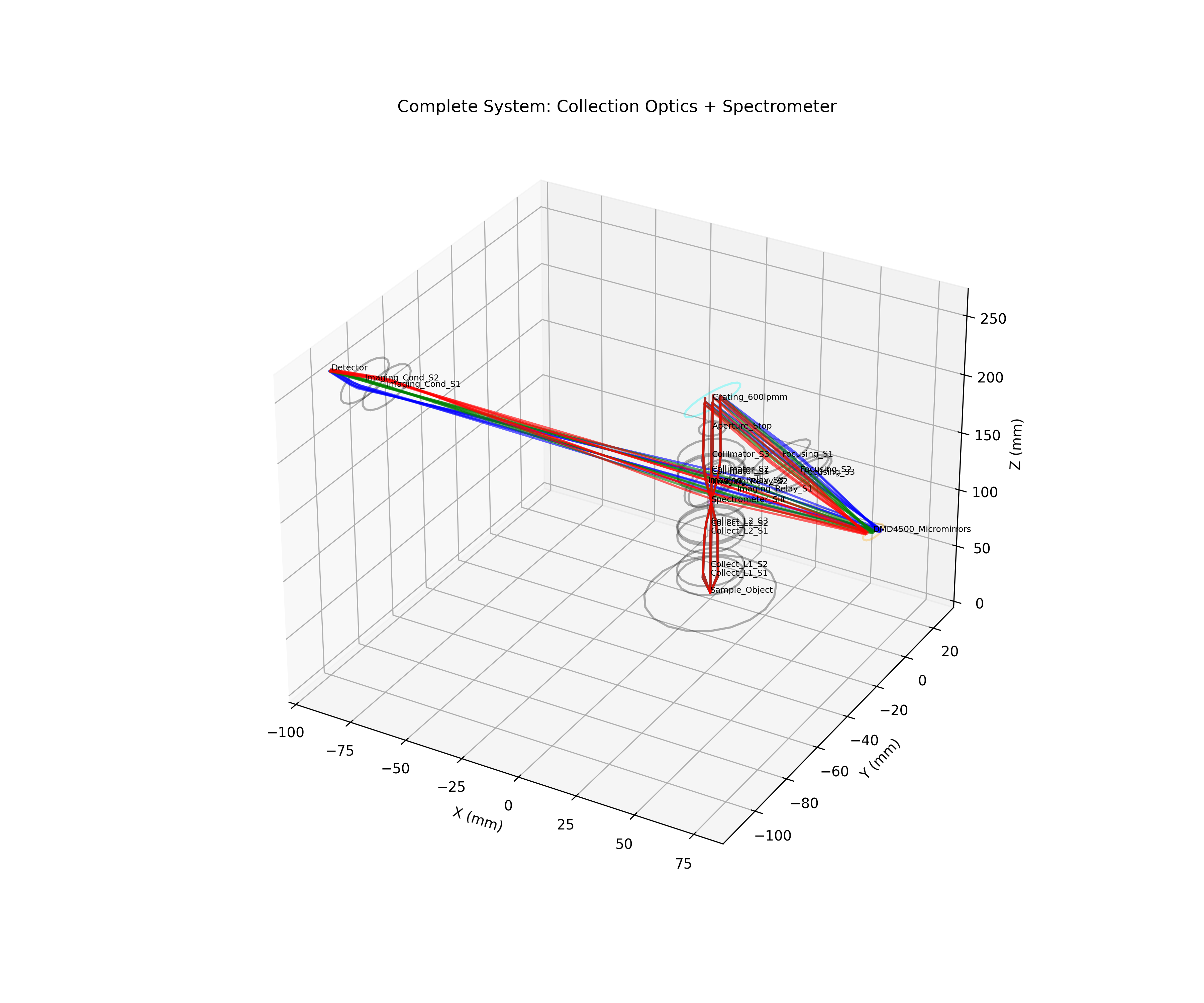
3D ray trace of a complete optical system
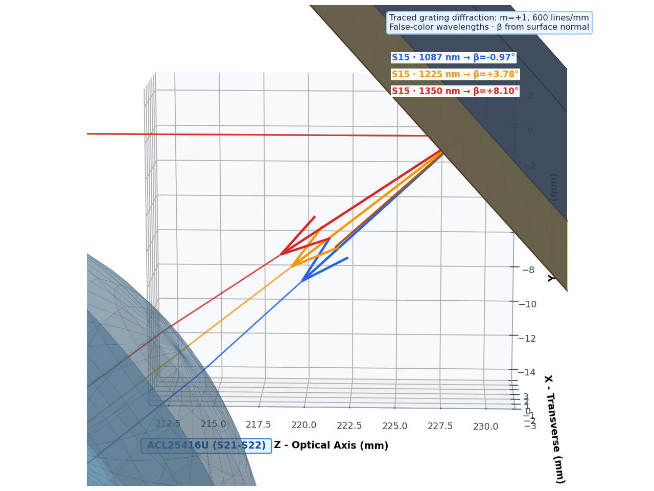
Grating diffraction in a NIR Raman spectrometer
Folded 1064 nm refractive spectrometer layout
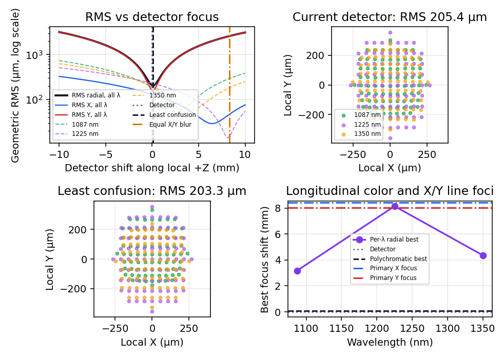
Best-focus analysis across the Raman band
All-mirror NIR spectrometer layout
3D-printable casing for the DMD Raman spectrometer
Included design presets (Herbert Gross handbook, Olympus, Thorlabs)
Cooke triplet 150 mm f/5.6Zeiss TessarDouble Gauss 50 mm f/2Petzval portraitPlössl, Erfle & Nagler eyepiecesSchmidt-Cassegrain & Ritchey-ChrétienOffner & Dyson relaysHigh-NA water multiphoton objectiveOlympus 25× NA 1.05 SRS microscopeTi:Sapphire Z-fold cavityFly's-eye & light-pipe homogenizersMichelson & Mach-Zehnder interferometers
Scientific Engineering Python · C++ · WebGL/Canvas

Custom Application & Instrument Development

Hardware-in-the-loop control, real-time spectroscopy & interactive physics

We build modular scientific applications connecting optical hardware (SLMs, deformable phase plates, galvo scanners, CCD/InGaAs spectrometers) with real-time signal processing and interactive browser simulations.

Closed-Loop AO Control
Zernike & DASH wavefront optimization algorithms for SLMs.
Spectroscopy Pipelines
Automated NIST calibration, baseline removal & ML classification.
Interactive Web Labs
Embeddable HTML5 Canvas optics & pulse simulators for articles.
Explore Interactive Simulations Lab → blog.py new "Title" --category simulation
⚡ Open Interactive Simulations Lab →

Loading article, figures and interactive scripts…

Article

Author
Juan David Muñoz-Bolaños
Biophotonics, Adaptive Optics & Computational Microscopy
Permalink
Interactive Simulations → SpMap & 2D Optics →
Press ESC to close Live Knowledge & Simulation Index
Advanced Imaging, Adaptive Optics & Lens Design
Juan David Muñoz-Bolaños · Leibniz Institute of Photonic Technology
Simulations Research & Thesis Microscopes Theory SpectraMap (GitHub) 2D Optics (GitHub) jmunozbolanos@gmail.com